Hello everyone, how are you all? i hope you all are doing well and i am too. Today i would like to tell about trichomonas vaginalis and what its significance in diagnosing it and how to diagnose it. First of what test is to be done?. PAP smear is done in females and we know that PAP smear is a screening test to rule out malignancy cervical cancer. Its a simple procedure and young age to old age will be done. After doing PAP smear by obstetrics and gynecology doctors they will send the pap smear with written form and labelling to pathology department. So whenever we receive the PAP smear we will check whether the name and details of the patient written on the form are correct or not and if any queries we will ask the obstetrics and gynecology doctors. After checking all details and then we will do staining for the slides and and it will take 2 hrs for staining, drying and mounting of all batches to make them ready/ These slides are submitted to us by technicians with labelling on the slide with lab number. Then we will again recheck and verify that the label number written on on the form is matching with the lab number written on the slide. We will start seeing the slide and today we are going to see how trichomonas looks like. Before that few things we have to know that as it is sexually transmitted infection females will be most commonly affected and males won't show any clinical symptoms and only females show clinical symptoms like vaginal discharge which is yellow-green, foul odour and itching. After hearing all these clinical symptoms, per vaginal examination is done and PAP smear is taken. Causative agent -Trichomonas vaginalis,single celled, flagellate and the most common pathogenic protozoan of humans and it is sexually transmitted infection. It causes trichomoniasis with the clinical features described before. Shape of protozoan - It is pear shaped with flagella and the nucleus is at one end not in the center. Microscopic picture -
10X maginification objective lens

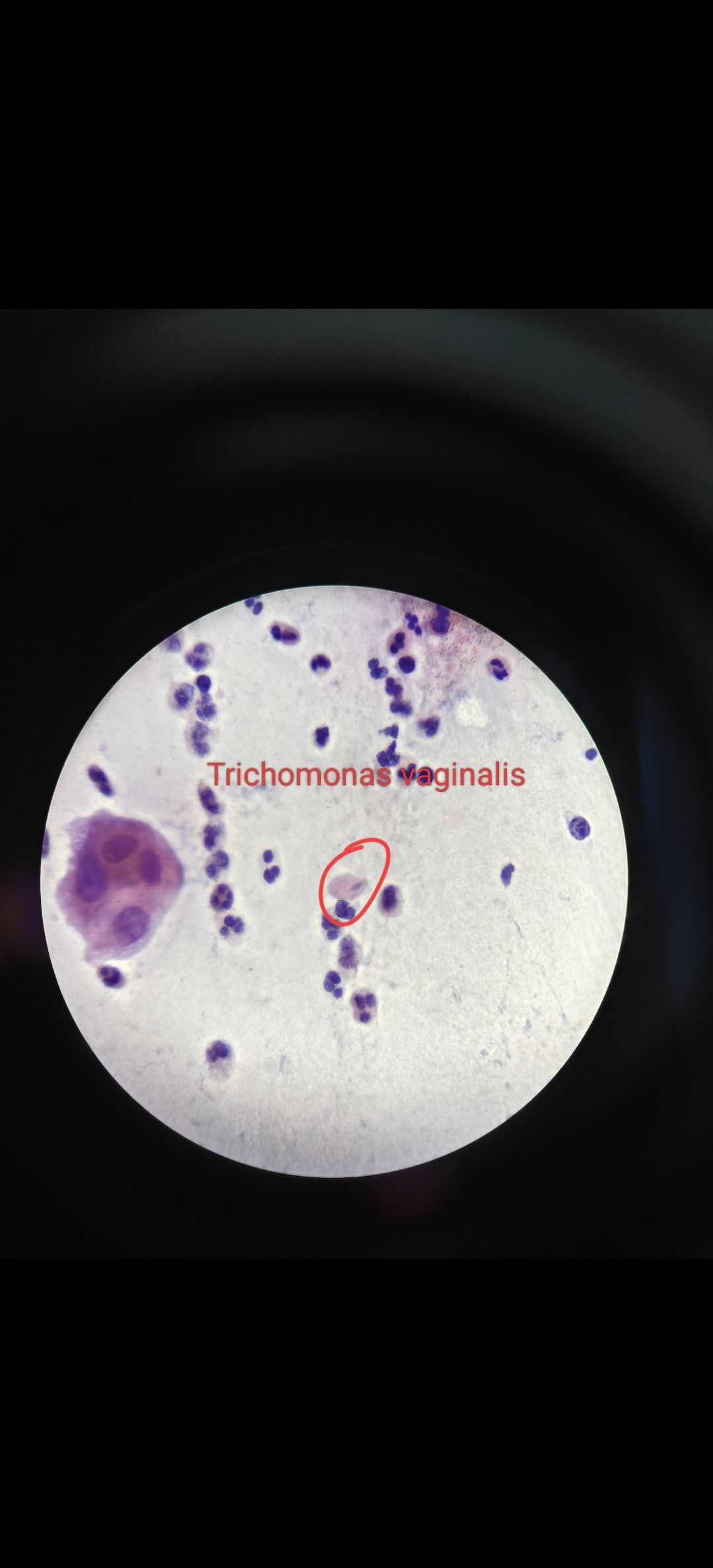 You can see in the above and below images that i have labelled trichomonas and see the shape of it which is pear shaped and nucleus is at one end and sometimes you can find that thin flagella if you carefully see it. This is how trichomonas is identified microscopically. So we will give report as trichomonas is present then the referring doctor will give treatment for the patient. The treatment for the patient is Metronidazole or tinidazole.
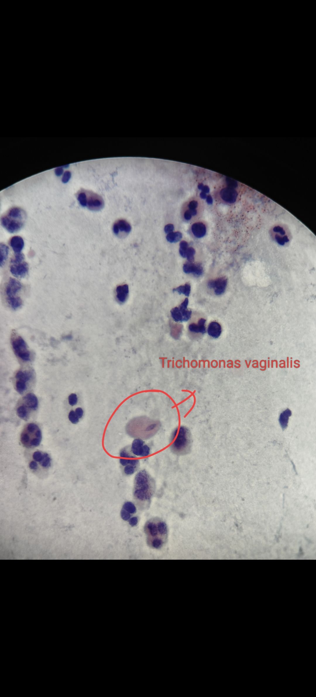
40X objective lens
I hope you understood well about trichomonas and its how important it is to diagnose it. We will come with new interesting topic stay tuned.
References
- The Bethesda System for Reporting Cervical Cytology, Ritu Nayar
Thanks for reading, with regards,